

Welcome to the Image Response Assessment Team Core Lab!
We are the Image Response and Assessment Team (IRAT) core lab of the Sydney Kimmell Comprehensive Cancer Center (SKCCC) in the Johns Hopkins School of Medicine.
Our role within the Cancer Center is to facilitate SKCCC member access to advanced medical imaging within their trials through:
- Protocol and Regulatory Guidance related to imaging
- Image Data Management
- Advanced Quantitative Image Analysis
Services
Protocol Development and Guidance
- Documentation Production and Review
- Imaging Protocol Development
- Weekly review of imaging on all protocols through the CRC Sub-committee forum
- Available to assist with IRB applications
- Radiation Exposure / Administration
- Risk Statements
Image Data
- Image Data Management
- Data De-Identification: The IRAT lab is an institutionally approved de-identifier of medical image data.
- Import/Export: The IRAT lab maintains a secure FTP server ("sFTP") which can facilitate the secure bulk exchange of study data with external collaborators.
- Archiving (trial specific): The IRAT maintains a partitionable research DICOM archive for the secure management of clinical trial imaging data.
Image Analysis
- Response Assessment Reports
- Response Assessment Criteria:
- PERCIST
- Lugano
- PCWG3 (Prostate Cancer Working Group 3)
- Curie
- Modalities:
- PET, PET/CT
- SPECT, SPECT/CT
- Bone Scan
We can work with your trial to provide modified versions of the above if required to comply with your sponsor reporting needs. In addition, we can also work with you to provide novel image analysis (e.g. Radiomic tumor metrics, Artificial Intelligence driven analysis) as your research may require.
- Quantitative Image Analysis
- Radiomics (Texture Analysis)
- Dynamic Analysis (Kinetic Modelling, Patlak Analysis)
- Artificial Intelligence (Automated lesion detection, early prognostic assessment)
Team IRAT!


Resources
Service request forms are available here:
Lab equipment available to approved users:
- PMOD - Kinetic Image Analysis Workstation
- MIM - PET/CT Image Analysis Workstation w/Neuro Analysis
- Mirada XD3 - PET/CT, SPECT/CT Fusion Image Analysis Workstation
- Nvidia DGX - AI Development/Training Workstation
Software can be found here:
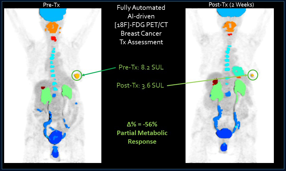
- Auto-PERCIST is a software application which implements and automates the algorithms and techniques described by the PERCIST v1.0 criteria for the quantitative analysis and response assessment determination of [18F]FDG PET imaging studies.
- Imager-4D is a multi-modal volumetric image analysis system designed for the review and quantitative analysis of radiological data. It includes tools for filtering, radiomic analysis and image labelling for machine learning. For dynamic datasets, in particular PET data, it includes integrated PATLAK modelling.
Auto-PERCIST
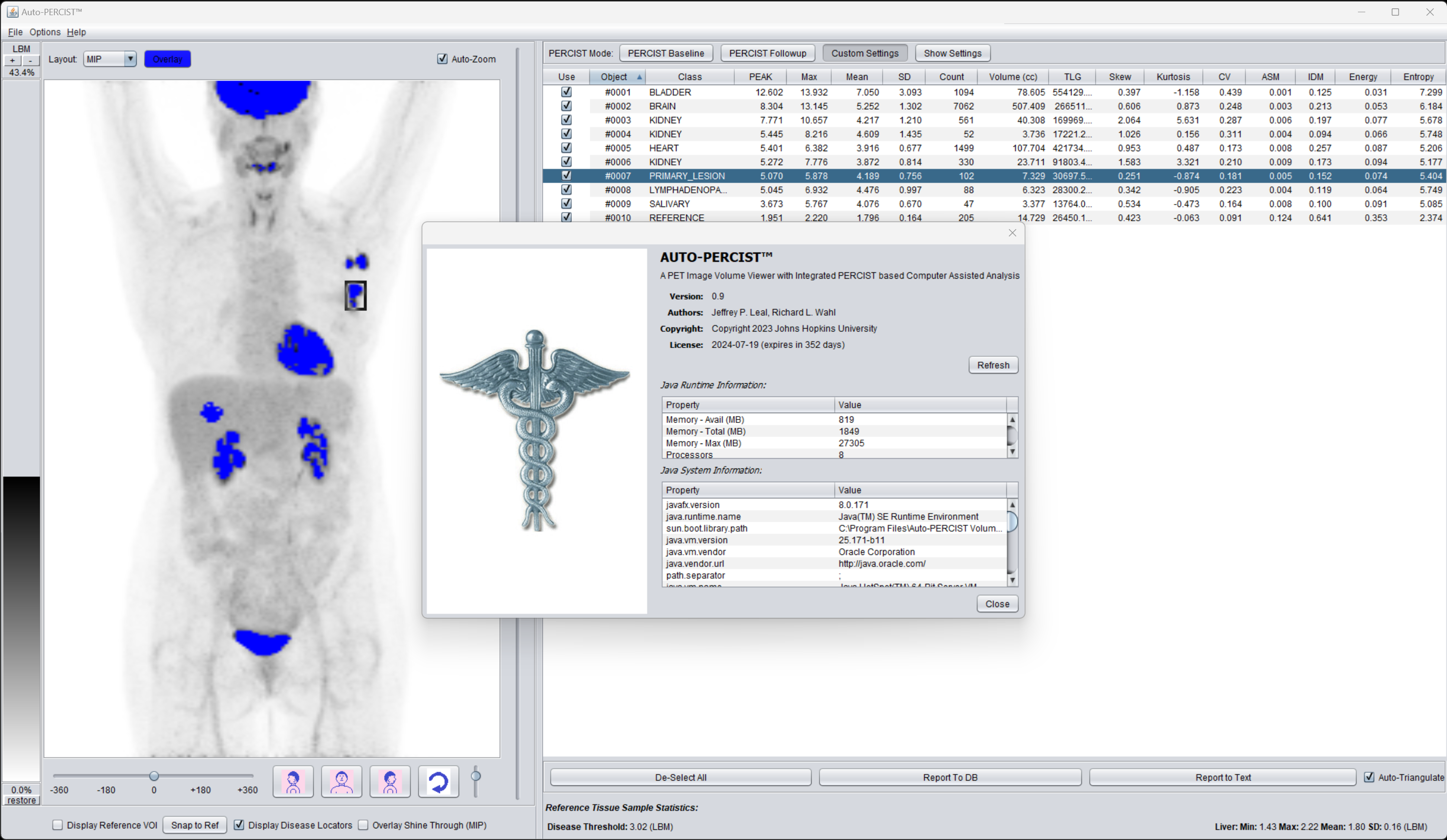
Auto-PERCIST is a software application which implements and automates the algorithms and techniques described by the PERCIST v1.0 criteria for the quantitative analysis and response assessment using [18F]FDG PET imaging. Through the implementation of these techniques, it performs an automated quantitative analysis of those regions of statistically significant elevated avidity, allowing the user to select those regions which are disease and storing the results of that analysis in database for comparison with future results and the automated determination of treatment response.
The software has been written in the Java language and is packaged to run on the Windows environment and integrate with freely available DICOM archives and SQL databases. Execution requires a license which is freely available to researchers.
The seminal paper describing the PERCIST v1.0 criteria can be found here: Some of the published work describing this software can be found here: We gratefully acknowledge funding from the National Cancer Institute P30CA006973. Please acknowledge the "Johns Hopkins Image Response Assessment Team (IRAT) Lab" and funding source when publishing work in which this software was used.
Imager-4D
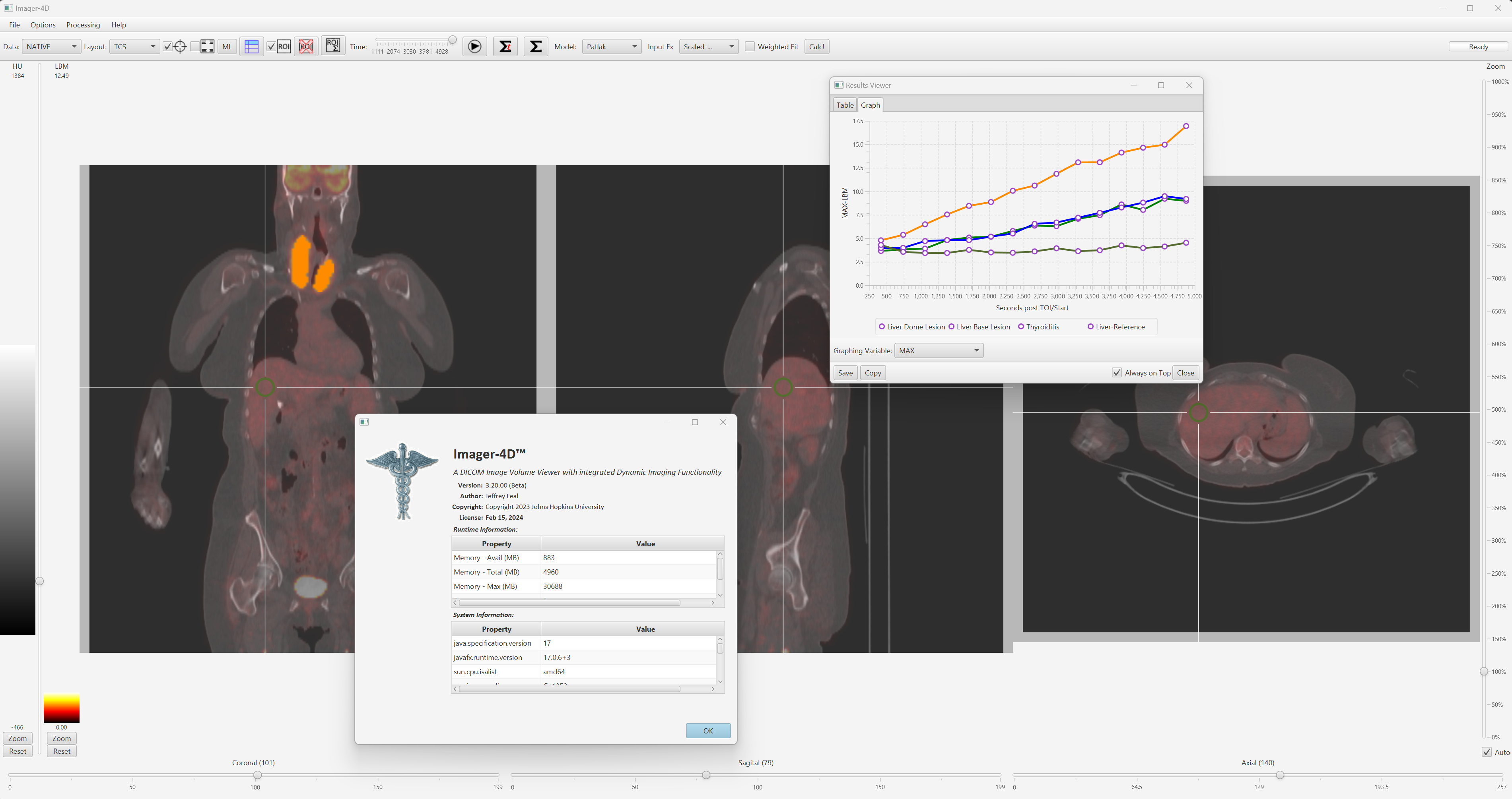
Imager-4D is a multi-modal volumetric image analysis system designed for the review and quantitative analysis of radiological data. With a focus on PET/CT (though able to work with all volumetric modalities), the application provides tools voxel normalization, image overlay, image filtering, volume of interest (VOI) analysis, as well as a host of other tools and features. In addition to the traditional quantitative tools, Imager-4D was designed from the ground up to work with 4-dimensional data (i.e., dynamically acquired volumetric datasets). Along with generating normalized Time-Activity-Curves (TAC), the system also has PATLAK modelling built-in. Finally, the system has advanced features which allow the user to easily generate training data for advanced machine learning systems.
The software was written in the Java language and is packaged to run on the Windows environment and integrate with freely available DICOM archives to simply data input. Execution using the base feature set is readily enabled with advanced features requiring a runtime license which is also freely available to researchers.
The original paper describing this software can be found here: We gratefully acknowledge funding from the National Institutes of Health EB024495 and the National Cancer Institute P30CA006973. Please include the above citation and funding acknowledgment when publishing work in which this software was used.
Contact
The IRAT lab is located at:
- Suite 300
- 550 North Broadway
- Baltimore, MD 21205
- email: iratlab@jhmi.edu
We are immediately adjacent to the Johns Hopkins Outpatient Center
Please note that in-person meetings in the IRAT are by appointment only. Please email a team member or the general lab email above to arrange an in-person meeting.